

 Freezing Procedures
With the ‘slow freezing technique’ used in the past, the viability and quality of the frozen eggs and sperm cells, and the embryos could be adversely affected during thawing. In today’s ‘fast freezing’ (vitrification) method, these cells and embryos maintain the same quality and viability after thawing.
Egg (Oocyte) Freezing
The process of freezing and storing healthy egg cells collected from the ovary in order for the woman to try and become pregnant in the future with use of fertility treatment is called ‘egg freezing’, also known as mature oocyte cryopreservation. In order to obtain eggs, egg cell enlargement injections used in IVF treatment are applied. The only difference is that after the eggs are collected, the good quality ones are frozen with the fast freezing (vitrification) method for than thawing them later on so they can be used in fertility treatment.
Egg freezing is recommended for the following patients:
- Patients who will undergo surgery for cancer
- Before cancer treatments such as chemotherapy, radiotherapy
- Before operations that may adversely affect ovarian function
- Those with a family history of early menopause
- Those whose ovarian reserve is found to be reduced by hormone tests and ultrasound
- In patients who cannot obtain live sperm cells in their partner on the day of egg collection in IVF treatment
Freezing Sperm Cells
We apply sperm freezing in the following cases:
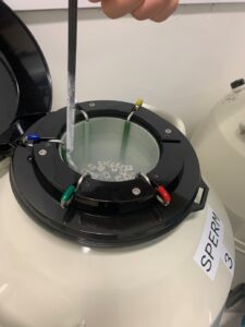 In patients who will undergo chemotherapy and radiotherapy due to cancer diagnosis, freezing can be performed in order to preserve reproductive functions.
- On the day of the procedure, if the person can not be physically present at the treatment site or will not be able to provide sperm sample due to psychological, socio-cultural or religious reasons, sperm sample given before can be frozen and stored.
- The leftover sperm obtained by MicroTESE (Microscopic Testicular Sperm Extraction) method during IVF can be frozen and stored for subsequent applications.
- Sperm can be cryopreserved to guarantee reproductive function before any kind of testicular surgery.
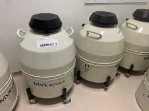
Embryo Freezing
The freezing and storage of good quality embryos obtained during the IVF procedure, sometimes due to medical necessity and sometimes at the request of the couple, is called ’embryo freezing’. Embryos can be frozen in the first 6 days after their development.
Who can undergo embryo freezing?
 In PCOS (Polycystic Ovary Syndrome), we prefer to freeze good quality embryos obtained through IVF treatment. This is because achieving pregnancy with embryo transfer in that treatment cycle can cause a very severe disease in the mother called ‘Ovarian Hyperstimulation’ (OHSS), which can lead to maternal death.
- In women who need to undergo surgery, chemotherapy or radiotherapy for cancer, embryos obtained by IVF are frozen and stored before the treatment.
- Sometimes more than the required number of good quality embryos can be obtained during IVF. In such cases, the good quality embryos remaining after the transfer are frozen.
- During IVF treatment, the chance of pregnancy decreases if the thickness of the inner lining of the uterus (endometrium) is below 7 mm at the time of embryo transfer. In such cases, the good quality embryos obtained are frozen, and in the following months, only treatment for improving the inner lining of the uterus for implantation is given before the embryos are transferred.
- Embryos biopsied for preimplantation genetic diagnosis (PGD) are frozen because the results of this analysis are available in a few weeks. According to the results, healthy embryos are thawed and transferred to the mother.
- If the woman’s ovarian reserve is decreasing and the family does not want to have children yet, embryo freezing is applied.
Preimplantation Genetic Diagnosis
Preimplantation Genetic Diagnosis (PGD) is a method that enables the selection of healthy embryos by genetic examination of the embryo before its transfer into the uterus in IVF treatment.
For this purpose, a biopsy of the embryo on day 5 is currently performed. One or two cells are taken from the embryo and used for genetic analysis. All chromosomes in the cell are analysed using the New Generation Sequencing (NGS) method. In pregnancies resulting from embryos that have undergone preimplantation genetic diagnosis, all screening tests during pregnancy follow-up must still be performed.
Preimplantation genetic diagnosis (PGD) is used in the following circumstances:
- Advanced maternal age (38 years and older)
- Severe male factor (severe oligospermia or azoospermia)
- One or both partners are carriers of a structural chromosomal disorder (translocation, inversion, deletion)
- History of recurrent pregnancy loss
- Recurrent IVF failure
- The family has a child with a chromosomal abnormality,
- Known carrier of a single gene disease or presence of a single gene disease in the family,
- Families’ desire to achieve a healthy pregnancy in a shorter time
Assisted Hatching
As the embryo develops, it is surrounded by a protective membrane. In order for pregnancy to occur, the embryo must naturally break free of this membrane and attach itself to the uterus. Sometimes embryos may have a thicker and harder membrane than normal for various reasons. In this case, it is thought to be useful to thin this membrane to facilitate the budding of the embryo before transfer.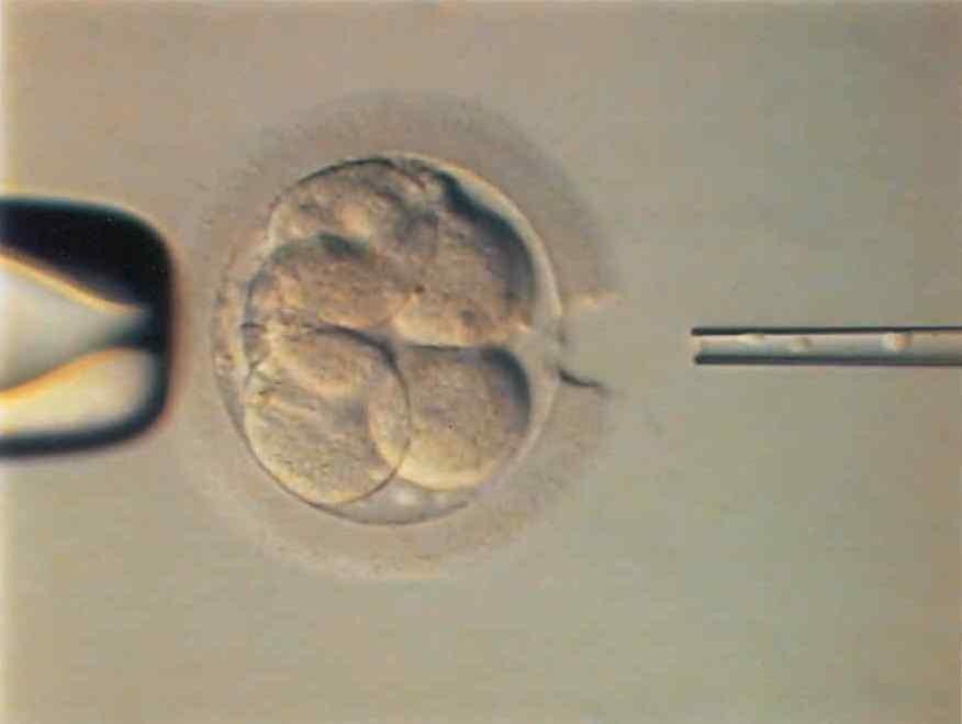 With this procedure, also called ‘assisted hatching’, the membrane is thinned in a certain area and the embryo is facilitated to bud from this point. We use laser technology for this procedure because it is much faster and safer for the embryo than other methods.
Who can undergo assisted hatching?
- Women aged 35 and over
- Three or more IVF failures
- In cases where the membrane around the embryo is excessively thick
- In cycles with frozen embryo transfer
Embryoscopy
In IVF, the most ideal conditions required for the development of the embryo from the time of embryo formation until the time of transfer are provided in the laboratory environment. The embryo is protected from external factors in a device called an incubator and its needs are met by the culture medium in which it is placed.
The embryo must be examined under a microscope in order to monitor their development and to determine the ideal transfer time. The ‘Continuous Embryo Monitoring System’ (embryoscope) has been developed to reduce the need to frequently take the embryo out of the incubator and examine it under a microscope.
The embryoscope is a safe incubation system used in IVF laboratories. With this technique, the development of each embryo is continuously recorded by a specially developed camera system. Thus, the embryo development follow-up process, which takes about 5 days, can be watched as an accelerated video image.
PRP (Platelet-Rich Plasma) Therapy
PRP application is the injection of plasma seperated from blood that is rich in platelets and growth factors for treatment of injured organs or tissues. It has been used in medicine for many years to help tissue repair in many areas other than gynaecology and obstetrics.
For PRP application, a quantity of blood taken from you is processed, the platelet- and growth factors-rich plasma part of the blood is separated and injected to the desired area for rejuvenation.
In the field of IVF, PRP application is used to rejuvenate the ovarian tissue and thicken the inner lining of the uterus (endometrium). The PRP injection to the ovary is performed under sedation anaesthesia, whereas if it is to be applied to the uterus, the patient is not given anaesthesia. The patient does not feel pain in both procedures.
Gynaecological Surgeries
Hysteroscopy
Hysteroscopy is the evaluation of the cervical canal and the uterine cavity using a lighted camera system. It is used for both diagnosis and treatment. Hysteroscopic operations are performed in the first few days following the end of menstruation. The areas of use of hysteroscopy in infertility are as follows:
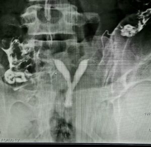 For both diagnosis and treatment of masses such as fibroids, polyps or intrauterine adhesions identified in hysterosalpingography (HSG).
- For the treatment of some congenital anomalies (intrauterine division, T-shaped uterus) detected in HSG, which may prevent pregnancy and may cause miscarriages.
- For the diagnostic evaluation of the uterus in women who have undergone three unsuccessful IVF treatments.
- For the treatment of adhesions which are due to previous miscarriage or intrauterine infections.
Laparoscopy
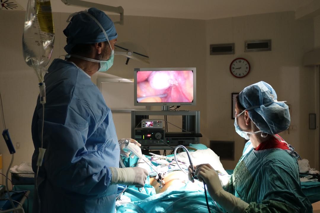
Laparoscopy is a modern surgical method in which the abdomen can be observed with a video camera system through a small incision made under the umbilicus (diagnostic laparoscopy) and when an operation is required, the necessary procedures can be performed with instruments applied through 0.5 – 1 cm. additional incisions made on the anterior abdominal wall (operative laparoscopy). The most important advantages of laparoscopy are that the scar is small, postoperative pain is less, recovery time is fast, hospital stay is short, and the risk of adhesion development betwwen abdominal tissues and organs after surgery is low.
The main areas of use for laparoscopic surgery in infertility are as follows:
- Diagnostic purposes
- Evaluation of uterus, tubes, ovaries
- Definitive diagnosis of endometriosis
- Elimination of tubal pathologies such as hydrosalpinx, ectopic pregnancy
- Opening of adhesions involving the ovaries, tubes and uterus
- Removal of endometrioma (chocolate cyst) and other ovarian cysts
- Correction of ovarian torsion (ovarian rotation around its own axis)
Microscopic (Microdissection) Testicular Sperm Extraction (Micro TESE)
Micro TESE is a surgical procedure performed under general anaesthesia in the operating room using an operative microscope to obtain sperm for IVF/ICSI applications. This operation takes approximately 1-2 hours. This method is more successful than the previously used standard testicular biopsy techniques in terms of finding live sperm in the testes of men who have no sperm in their semen (azoospermia) due to reasons not related to obstruction of the sperm-conducting ducts. If there are any leftover sperm, these can also be frozen.
Leave a Reply
You must be logged in to post a comment.